
GA2-57
Airway Management in Infant with Acute Respiratory Distress from Cystic Lymphadenitis
Chu M, Ekeoduru R, Wadhwa N, Roy S
University of Texas Health Science Center at Houston, Houston, TX, USA
Intro
Facial mass causing respiratory distress is a pediatric emergency. These children are known difficult airways making anesthesia preparedness critical. The ASA has not published the prevalence of facial mass complicating anesthetic management, but in the UK, 40% of reported cases in the NAP4 audit (a major UK study on complications of anesthesia) had head and neck pathology.1 Our case describes successful airway management of an infant with acute ventilatory failure from a large, cystic facial mass.
Case
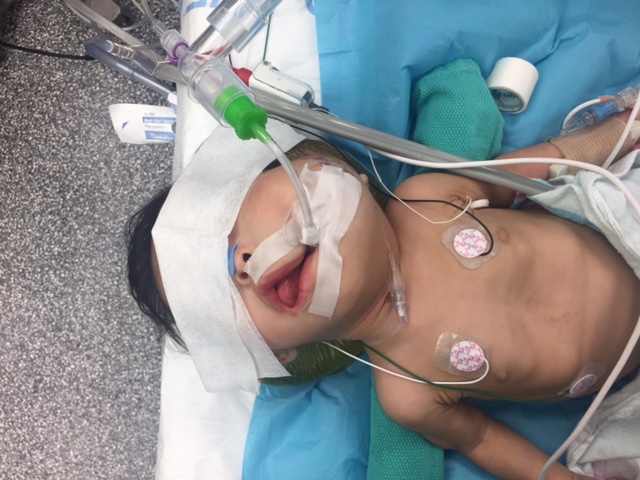
A 7 month old girl with a large left facial mass presented with hypoxia and respiratory distress. The mass was determined to be cystic lymphadenitis via imaging. The child was mid-flight, when she decompensated with facial pallor. The flight was diverted and she was transported to our facility.
There was a large, ill-defined mass over the left mandible & neck to the shoulder. Flexible laryngoscopy was done by ENT at bedside, but they were unable to visualize airway structures. Afterwards, the child had worsening retractions and desaturations. She was emergently transported to the OR for airway protection.
She was induced with titration of Sevoflurane and 100% FiO2 while maintaining spontaneous respiration. A nasopharyngeal airway was needed for airway patency. Versed & Precedex were titrated for adequate anesthetic depth. Direct laryngoscopy and rigid bronchoscopy were done by the surgeons.
Initially, it was difficult to visualize the larynx because the tumor filled the oropharynx and tongue base. With manipulation, the cords were observed deviated laterally secondary to the mass. With a view, a 3.0 cuffed ETT was placed. Propofol was then given to increase depth of anesthesia for the controlled tracheostomy. Postop, she was weaned to trach collar on RA and discharged POD 20.
Discussion
Facial mass can be congenital or acquired. Lymphadenitis is the most common cause.2 Airway compromise is a concern as the mass has the potential for airway obstruction that can make mask ventilation and intubation difficult. These patients may be stable initially, but they can decompensate unpredictably and quickly. In our case, a team effort between ENT and pediatric anesthesia was crucial. The primary goal was to maintain spontaneous respiration while achieving a deep anesthetic plane to allow for airway evaluation. This can be achieved by titrating Sevoflurane with a combination of Precedex, Ketamine and/or Versed. Establishing an emergent surgical can be difficult which is why the maintenance of spontaneous ventilation is so important.
References:
1. Morosan M, et al. “Anaesthesia and common oral and maxiollo-facial emergencies.†Continuing Education in Anaesthesia Critical Care & Pain, VOl 12, Iss 5, 1 Oct 2012, 257-262
2. Khanna G, et al. “Causes of Facial Swelling in Pediatric Patients: Correlation of Clinical and Radiologic Findings.†RSNA. Jan-Feb 2006. Vol 25, Issue 1
-
 GA2-57 Image 1
Top


