
CR1-181
Ultrasound-guided, continuous brachial plexus blockade in a neonate with upper extremity gangrene
Hubbard R, Cappuccio E, Martin D, Dairo O, Smith T, Bhalla T, Corridore M, D J
Nationwide Children's Hospital, Columbus, Ohio, USA
INTRODUCTION: Brachial plexus blockade is a well-reported regional anesthetic technique in children [1]. Despite its efficacy, this block is infrequently used in the younger pediatric population [1]. Indeed, only two case reports document single-shot, landmark-based, axillary brachial plexus blocks in neonates [2,3]. Catheter-based continuous peripheral nerve blocks have been reported only in children over 1 year of age [4]. We report the ultrasound-guided placement of a continuous brachial plexus catheter in a neonate born with right upper extremity gangrene, requiring serial debridements.
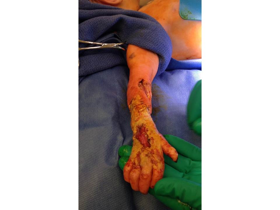
METHODS: A 10-day-old, 3.4kg, previously term female with a history of intrauterine gangrene to the right upper extremity (RUE) presented for operative debridement. Serial wound vacuum changes were planned. To reduce repeated exposure to anesthetic agents and treat postoperative pain, a supraclavicular nerve catheter with continuous local anesthetic infusion was planned. Following the induction of anesthesia and endotracheal intubation, the block site was cleansed with betadine. Ultrasound (US) imaging was used to identify the brachial plexus. A 20-gauge, 1.5 cm angiocatheter was inserted above the clavicle, with the needle tip being visualized on US using an in-plane view. Once placed in proximity to the brachial plexus, the needle was removed, leaving the angiocatheter at a depth of 1 cm. Catheter tubing was attached and a clear dressing applied. A bolus dose of 3mL of 0.2% Ropivicaine was injected. Postoperatively, an infusion of chloroprocaine 1.5% at 2 mL/hr was started.
RESULTS: The local anesthetic infusion was continued for 7 days, during which time the patient required only occasional supplemental doses of opioids. She underwent dressing changes on post-operative days 3 and 6. In both cases0.5% ropivicaine (between 1.0-1.2mL) was bolused through the catheter for pain control, and the patient tolerated dressing changes at the bedside. The catheter was removed on postoperative day (POD) 7. Subsequent dressing changes (POD 13) required the use of general anesthesia. Following successful skin grafting, the pateint was discharged home on day of life 26.
DISCUSSION: With increasing concern for the potential neurocognitive impact of anesthetic agents in early life, potential alternatives must be sought. This is especially true for children whose pathologies require repeated procedures. In our case, the successful placement of a brachial plexus catheter spared the patient two additional anesthetic exposures, with the added benefit of excellent pain control. To our knowledge, this is the first documented use of this technique in neonates.
REFERENCES
1.Tobias JD. Paed Anesth. 2001;11:265-75.
2.Masseri A. Paed Anesth. 2000;10:527-530.
3.Breschan C. Paed Anesth. 2004;14:681-84
4.Dadure C. Anesth Analg. 2003;97:687-90.
-
 CR1-181 Image 1
Top


