
GA1-53
TRACTION RELATED CHANGES IN EVOKED POTENTIALS DURING SCOLIOSIS CORRECTION – A retrospective review
1Lutwin-Kawalec M, 1Choudhry D, 2Brenn B, 1Sacks K
1Nemours/AIDHC, Wilmington, DE, USA; 2Vanderbilt University, Nashville, TN, USA
Background: MEPs and SSEPs monitoring has become a standard of care for posterior spinal fusion surgery (PSF). Traction is frequently used during PSF with severe scoliosis. We report a series of cases where neurophysiologic changes were observed with cranio-femoral (CFT) traction. Aim of the study was to evaluate various factors that may be of relevance for occurrence of changes in TcMEPs whilst CFT is applied during PSF. If changes were observed in TcMEPs, the measures effective in restoring the potentials were also evaluated.
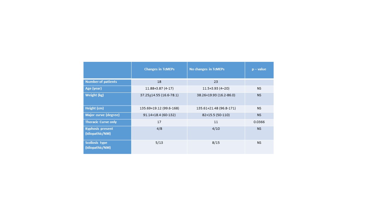
Methods: Following IRB approval, data was collected retrospectively for all children, between June 2011 and June 2017 who had CFT applied during PSF surgery. The same surgeon performed all surgeries and children were divided in two groups: Group-1: Changes seen in TcMEPs with traction – 18; Group-2: No changes seen in TcMEPs with traction – 23.
Following routine monitors placement and inhalational induction, IV lines were secured, ETT was placed and invasive BP monitoring was established. Total intravenous anesthesia (TIVA) was then commenced and monitoring was established. Tongs were applied to the skull; children were then turned prone and placed in Jackson frame. Boot traction was applied to lower extremities (up to half the body weight) to reduce the deformity. TcMEPs were obtained at baseline, after traction application and during the prep phase.
Following parameters were recorded: demographic data, type of scoliosis (idiopathic/neuromuscular), location and degree of major curve (thoracic/lumbar), presence of kyphosis and changes in evoked potentials (SSEP, TcMEPs). If changes in potentials were observed, effective interventions were also recorded.
Nominal data was analyzed with chi-square test and interval data analyzed using independent sample t-test. Significance was assumed at p<0.05.
Results: Both groups were similar in demographics (Table-1). No difference was observed in type of scoliosis, severity of major curve or presence of kyphosis. Significantly more children with thoracic curve belonged to the group who had changes in TcMEPs (p<0.05).
Following three steps were taken to restore potentials: Reduction/removal of traction, increase in MAP and hardware manipulation. One intervention-6 patients; 2 interevntions-8 and 3 interventions-4. TcMEPs could be successfully restored in all children.
Discussion: TcMEPs are very sensitive to spinal cord ischemia during PSF. Our retrospective evaluation showed that likelihood of changes in TcMEPs whilst intraoperative CFT is applied during PSF is as high as 44%. Correlation between thoracic curve and changes in TcMEPs was seen. Neuromonitoring must be started prior to CFT, TIVA established soon after induction and muscle relaxant avoided for prompt recognition of changes in TcMEPs. Prompt reduction/removal of traction and increases in MAP are necessary to restore potentials.
Reference: Lewis, SJ et al. Spine (2011) 36 (20):1627-38
-
 GA1-53 Image 1
Top


