
NM-370
Does This Look Normal To You? The Importance of Recognizing Ultrasonographic Neonatal Spine Variation for Caudal Epidural Placement: A Case Report
Nia S, Falciola V, Gomez-Mourad A
Boston Children's Hospital, Boston, MA, USA
Introduction
Ultrasonography for regional interventions has revolutionized the practice of pediatric anesthesiology and pain medicine.This modality has increased confidence in utilizing interventions to treat pain in this vulnerable population, however, this innovation makes for challenges unique to neonates. With advanced imaging, issues are encountered in recognizing anatomic features which are specific to normal development and growth. In this case report, a caudal epidural injection under real-time ultrasonography revealed unusual images of a prominent filum terminale enhanced by a lack of bone shadowing due to immature non-ossified vertebrae. This is a normal finding seen in neonates with incomplete bone calcification. These results raised questions as to whether other practitioners are prepared to interpret such findings. This case report underscores the importance of being prepared to encounter anatomic differences in neonates which may lead a less astute practitioner to abandon an otherwise beneficial procedure under an erroneous presupposition.
Methods
A 2.7kg male former 26 week infant (corrected gestational age 41 weeks) was scheduled for an inguinal herniorrhaphy. The patient’s medical history was significant for birth weight of 850g with APGAR scores of 5/7 followed by respiratory complications requiring prolonged mechanical ventilation.
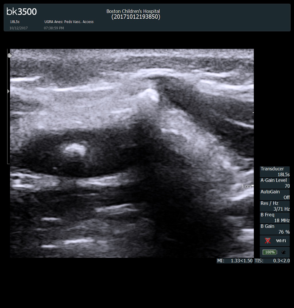
Given his hospital course and medical history, the intraoperative anesthetic plan was for a caudal epidural catheter infusion of 3% chlorprocaine with IV dexmedetomidine infusion. The caudal catheter was to be placed under real-time ultrasonography using a BK3500 machine (BK Medical, Peabody MA USA). Upon scanning the sacrum and coccyx, no cutaneous markers of dysraphism were encountered including hair tuft, or skin dimpling. Unconventional ultrasound findings were encountered most noticeably in the transverse plane which were recognized by the pediatric regional team as a prominent filum terminale, a normal variant. The caudal epidural catheter was placed uneventfully and the case proceeded successfully; the patient was discharged home thereafter.
Discussion
With current practices trending towards the use of ultrasonography for regional procedures, it is essential that interventionalists be aware of physiologic variations that are unique to neonates. Pediatric regional anesthesiology is at the forefront of point-of-care procedural ultrasonography and as such, it behooves practitioners to familiarize themselves with these essential variations so as not to mistakenly diagnose an incidental pathologic finding and unnecessarily withhold treatment. This case report aims to raise awareness of this crucial matter to ensure a successful, safe pediatric regional practice.
Conclusion
It is essential for regionalists to familiarize themselves with variants in neonatal spinal ultrasonography for pain procedures to ensure the safety and efficacy of the intended procedure.
-
 NM-370 Image 1
Top


