
GA3-76
Quantitative MRI Measures for Infant Brain Growth: Prematurity as a Risk Factor in the Setting of Surgery and Critical Care with Prolonged Sedation
1Goins S, 2Mongerson C, 3Jennings R, 3Grant P, 3Bajic D
1Middlebury College, Middlebury, Vermont, USA; 2Center for Pain and the Brain (P.A.I.N.) Group, Boston, MA, USA; 3Boston Children's Hospital, Boston, MA, USA
Introduction: Opioids and benzodiazepines are routinely used for pain and sedation management when treating critically ill infants. It is known that prolonged use of these drugs is associated with drug tolerance and dependence, but our understanding of their effects on brain development is limited3. We hypothesized that infants exposed to prolonged sedation will have decreased brain diameters and increased fluid-filled spaces compared to healthy, naïve infants, suggestive of delayed brain growth.
Methods: T1-weighted MRIs from 21 patients (full-term N = 8; preterm N = 13) from Boston Children’s Hospital who underwent surgical repair for long-gap esophageal atresia and 18 healthy infants were included. Corrected age at scan for preterm infants was calculated as follows: chronological age in weeks–[40 weeks–gestational age at birth in weeks]. Following manual orientation using Freeview, 4 brain diameters were measured by two blinded researchers using ITK-Snap[1,2]: interhemispheric distance, brain and bone biparietal diameters, and biventricular width. Calculated end-point analyses were: (1) biparietal diameter difference (bone - brain biparietal diameter), (2) ventricular-brain ratio (biventricular width / brain biparietal diameter), and (3) interhemispheric-brain ratio (interhemispheric diameter / brain biparietal diameter). Normality was assessed using the Shapiro-Wilk test. Pearson and Spearman correlations were used to study the effects of post-natal age on brain diameters as appropriate. Kruskal-Wallis and Mann-Whitney tests were used to study the effects of group on ventricular-brain and interhemispheric-brain ratios.
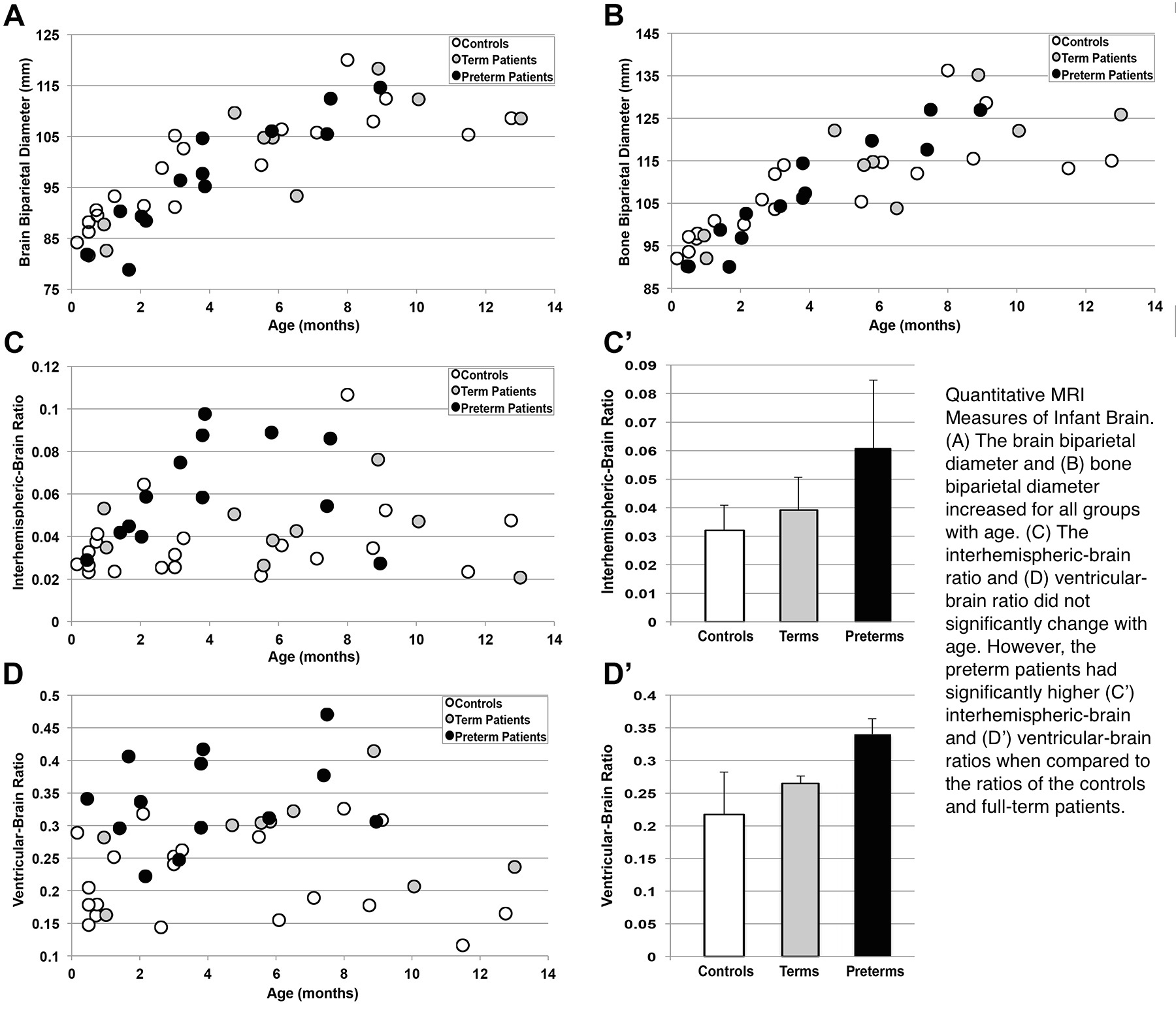
Results: Interobserver reliabilities from 42 brain MRI scans as measured by two independent researches were very high. While the brain and bone biparietal diameters significantly increased with age for all three groups, the interhemispheric-brain and ventricular-brain ratios showed a weak correlation with age. The interhemispheric-brain and ventricular-brain ratios of the preterms were significantly higher than those of the controls and full-terms.
Discussion: Brain and bone biparietal diameters increase with age for all groups. The preterm patients have significantly larger interhemispheric-brain and ventricular-brain ratios, suggestive of cerebrospinal fluid increase regardless of age.
Conclusions: Quantitative brain measures may be used as a predictive tool to help identify patients at risk of delayed brain growth.
References
1. Kidokoro H. et al. New MR imaging assessment tool to define brain abnormalities in very preterm infants at term. AJNR Am J Neurorad, 2013. 34(11): p. 2208-14.
2. Maunu, J., et al., Ventricular dilatation in relation to outcome at 2 years of age in very preterm infants: a prospective Finnish cohort study. Dev Med Child Neurol, 2011. 53(1): p. 48-54.
3. Anand, K.J., et al., Tolerance and Withdrawal From Prolonged Opioid Use in Critically Ill Children. Pediat, 2010. 125: p. 1208-1225.
-
 GA3-76 Image 1
Top


