
CA-40
Intraoperative isoflurane exposure predicts reduced frontal lobe connectivity compared to dexmedetomidine in neonates with congenital heart disease
1Adams P, 2Lee V, 2Meyers B, 3Dennis L, 1Beluk N, 1Baust T, 1Saenz L, 1Domnina Y, 1Sanchez-de-Toledo J, 2Schmithorst V, 1Panigrahy A
1Children's Hospital of Pittsburgh of UPMC, Pittsburgh, PA, United states; 2University of Pittsburgh School of Medicine, Pittsburgh, PA, United states; 3Regent University, Pittsburgh, PA, USA
Introduction: The GAS and PANDA studies showed a single anesthetic exposure of short duration does not adversely affect neurocognitive outcomes in children [1-2], however the MASK study showed an association between multiple anesthetic exposures and decreases in cognitive ability [3]. Unfortunately, babies born with critical congenital heart disease (CHD) often require numerous, lengthy anesthetics during infancy. Interestingly, dexmedetomidine (DEX) mitigates neuronal death caused by other anesthetics [4]. This study examined the hypothesis that longer volatile anesthetic (ISO) duration and lower DEX/ISO ratio would result in impaired brain structural and functional connectivity as determined by postoperative functional MRI (fMRI).
Methods: Prospective observational study examining neonates with complex CHD requiring neonatal intervention. The effects from ISO duration (min), total ISO exposure (ml) (estimated as product of averages of VE, EtISO, and DurationISO), total DEX dose (mcg/kg), and DEX/ISO ratio during cardiac surgery were investigated using manual tractography (MT), voxel-based morphometry (VBM), diffusion tensor imaging (DTI), and resting blood-oxygen-level dependent (BOLD) postoperative fMRI modalities.
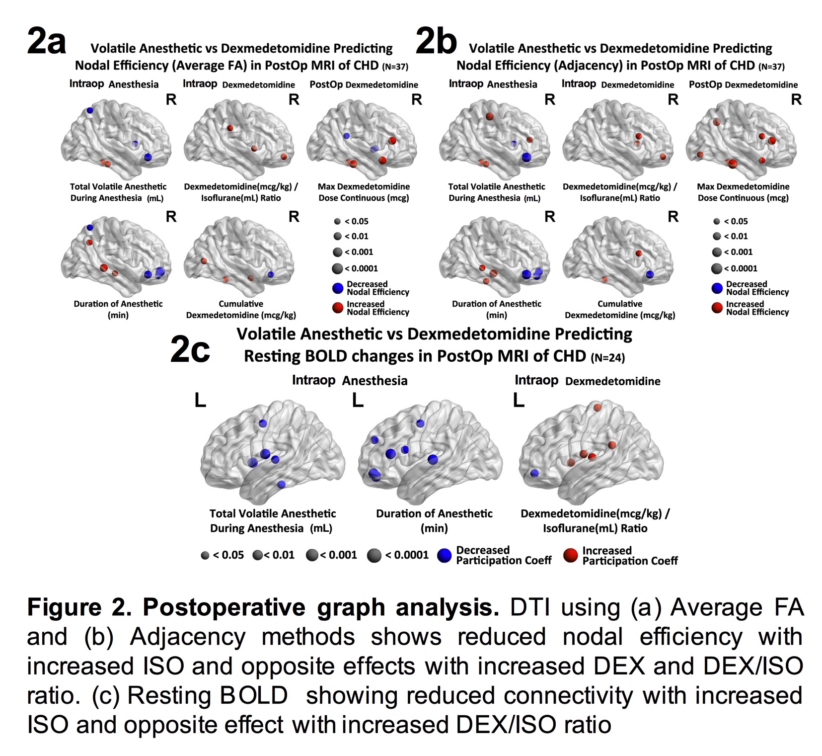
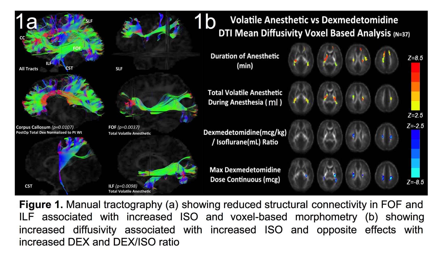
Results: 37 neonates underwent the study protocol. MT analysis showed higher total ISO dose was associated with reduced structural connectivity involving the fronto-occipital fasciculus (FOF) and the inferior longitudinal fasciculus (ILF) (Fig 1a). VBM diffusivity analysis showed both ISO duration and total ISO dose was predictive of increased mean diffusivity (opposite of normal developmental change) in frontal lobe regions that overlap with the FOF and ILF, while DEX had an opposite effect (Fig 1b). Graph analysis of DTI and resting BOLD showed that both ISO duration and total ISO dose were predictive of reduced nodal efficiency (Fig 2a-b) and reduced brain connectivity (network segregation) (Fig 2c) in the frontal lobe, respectively, while DEX had an opposite effect.
Discussion: Higher ISO exposure is associated with reduced frontal brain connectivity in CHD neonates using multiple fMRI approaches. In contrast, DEX exposure was associated with metrics of improved brain connectivity using the same analytical approaches. Given FOF and ILF abnormalities have been observed in patients with ADHD [5], our novel observations may help explain the behavioral abnormalities identified by Hu, et al [3]. Complex CHD patients often require numerous, lengthy anesthetics, suggesting all efforts for neuroprotection should be employed.
Conclusion: Higher DEX in relation to ISO exposure confers improved brain connectivity in CHD neonates.
-
 CA-40 Image 2
 -
 CA-40 Image 1
Top


