
AET-24
Laryngotracheal cleft in a neonate with complex congenital cardiac defects
Stamm K, Muellerleile P, Svendrowski M, Ahmad N
Saint Louis University, Saint Louis, Missouri, United states
Laryngotracheal clefts (LC), a congenital deformity of the posterior portion of the larynx and trachea, are a rare and often challenging congenital airway malformation. Occurring in every 10,000-20,000 live births, LCs cannot be diagnosed prenatally, and are usually found when symptoms manifest. We present a multi-staged case study of a neonate requiring surgical correction of cardiac anomalies with a laryngeal cleft that was diagnosed on initial intubation attempts.
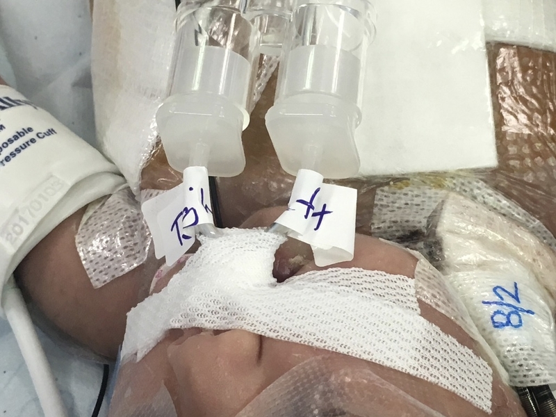
Our patient is a one day-old full term female with total anomalous pulmonary venous return diagnosed prenatally. She presented for repair of TAPVR on day one of life. She was induced and ventilated uneventfully. A 3.0 mm cuffed endotracheal tube was introduced into the trachea and appeared to be main-stemmed upon auscultation. When the ETT was withdrawn a large leak was noted. Further attempts produced similar results. A 3.0 mm ETT was then main-stemmed in order to ventilate the patient. An emergent bronchoscopy revealed a Type III laryngeal cleft. Due to the urgent need for cardiac repair and difficulty in ventilating, ENT performed a repair of the LC. The patient was transported to the ICU with a 3.0 mm uncuffed ETT in place. On day 9 of life, she underwent cardiac repair. Post-operatively, she was difficult to ventilate and her chest x-ray showed white-out of her left lung. Repeat bronchoscopy identified that her cleft repair had broken down due to prolonged intubation. She was placed on ECMO support due to ventilatory and hemodynamic instability. There was continued difficulty with endotracheal tube placement due to a deep cleft and short trachea so she returned to the OR for a bronchoscopy, while still on ECMO. Under direct visualization a 2.5 mm uncuffed ETT was placed in the right mainstem and another was placed in the left mainstem. A Y-adaptor was connected to both tubes and both lungs were ventilated.
Laryngeal clefts have been characterized by a modified Benjamin and Ingles scale into five types (O-IV) ranging from a submucosal cleft (O) to a cleft which may extend to the carina (IV). With type III, or IV LCs, surgical repair is often complicated, requiring multiple revisions, with a high rate of morbidity and mortality. Proper ventilation is of utmost importance in these patients, to prevent aspiration and pulmonary complications. If significant respiratory distress necessitates endotracheal intubation, then great care should be taken when placing the ETT, as proper placement can be difficult. Bronchoscopic guidance should be used to guide positioning. Early extubation is encouraged in repaired LCs because prolonged intubation can delay healing of the repair site.
Sonmez, K. et al. Our experience in two cases of type IV laryngotracheoesophageal cleft with a diagnosis of antenatal esophageal atresia. The Pan African Medical Journal, 2017; 26: 55.
Leboulanger, N. et al. Laryngo-tracheo-oesophageal clefts. Orphanet Journal of Rare Diseases. 2011; 6: 81.
-
 AET-24 Image 1
Top


