
NM-383
A Retained Arterial Catheter Fragment Migrates to Base of Thumb and Requires Two Subsequent Surgeries for Removal
FitzGerald K, Waller E
University of North Carolina at Chapel Hill, Chapel Hill, NC, United states
Introduction:
Placement of a radial arterial line for hemodynamic monitoring or serial lab draws is frequently performed during the perioperative period. We present an unusual but serious complication of a retained catheter fragment in the radial artery (RA), requiring surgical intervention.
Case report:
A 10 y/o female with PMH significant for heterotaxy syndrome, AVSD s/p repair, and complete heart block presented for removal and replacement of pacemaker. Induction, intubation, and PIV placement went smoothly. An arterial line was placed due to repeat thoracotomy. The left RA was cannulated on first attempt using a 22-gauge PIV catheter. The surgery was uneventful, and the pt was extubated and admitted to the PICU.
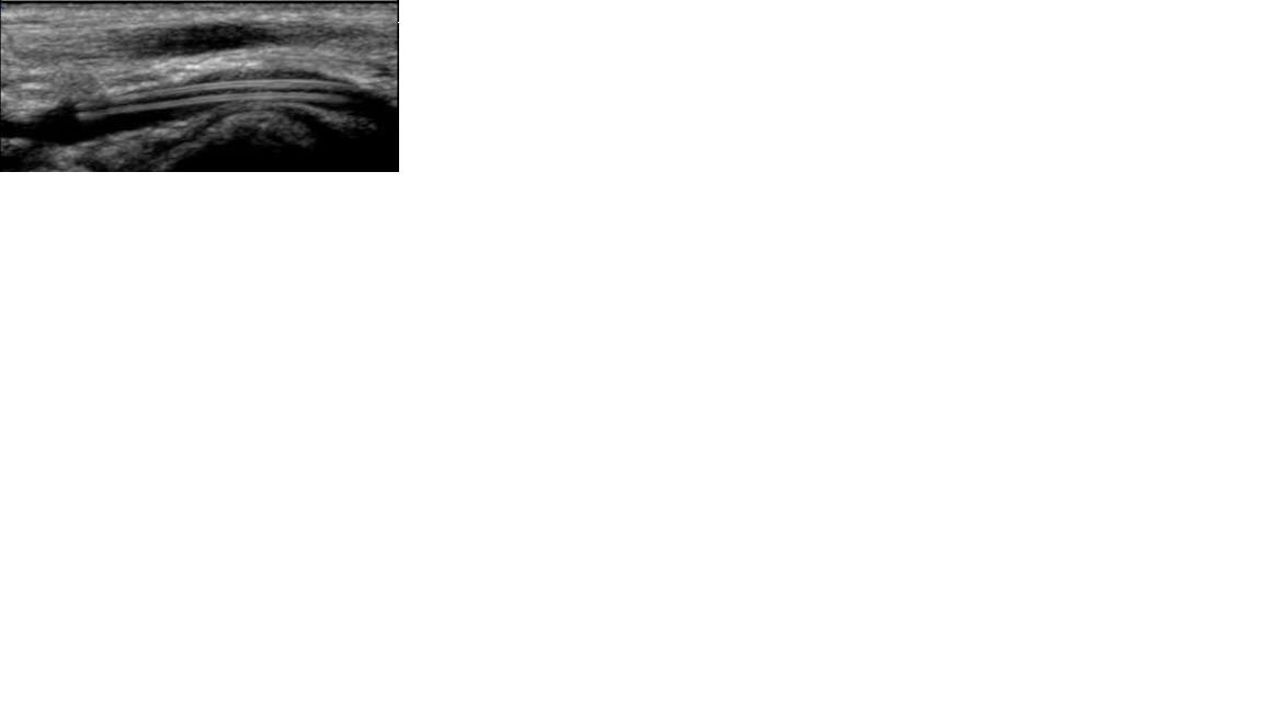
On POD1, the arterial line was removed and it was noted that ~2 cm of catheter was missing. The catheter fragment was easily palpated in the wrist and visualized on bedside ultrasound by the PICU fellow. The pt was taken to the OR later that day by vascular surgery for catheter retrieval under GA; however, the surgical team was unable to locate the fragment after dissection down to the RA. Angiography showed no filling defect, and the procedure was aborted.
Formal vascular studies obtained on POD2 demonstrated migration of the fragment distally to the base of the thumb. Orthopedic surgery was consulted, and the pt underwent hand exploration under GA on POD3. The fragment was found to have migrated further to the common digital artery (CDA) of the index finger. After difficult dissection, the fragment was removed intact and repair of the CDA and RA was performed. Post-op, the pt had mild paresthesias of the left index finger and abnormal two-point discrimination. This was thought to represent a neuropraxia injury that would resolve with time.
Discussion:
Complications of arterial cannulation include distal ischemia, hematoma formation, infection, thrombosis and embolism, and retention of catheter fragment (1). Causes of catheter damage include shearing by stylets during placement, accidental cutting with scissors during removal, and mechanical stress due to repeated movement (2, 3). In this case, there was no obvious external cause for damage to the catheter. Notably, a 22-gauge PIV catheter meant for low-pressure venous access was used in place of a prepackaged arterial cannula meant for higher pressures. This, plus mechanical stress, may have been enough to cause breakage.
This case demonstrates the importance of checking all arterial catheters upon placement and removal, and highlights a potentially serious risk of the practice of using venous catheters as arterial lines in children.
References:
1. Durie M, et al. Incidents relating to arterial cannulation as identified in 7525 reports submitted to the Australian incident monitoring study. Anaes and Intensive Care 2002;30:60–5.
2. Moody C, et al. Ultrasound guided location and removal of retained arterial cannula fragment. Anaesthesia 2009;64:338–9.
-
 NM-383 Image 1
Top


