
NM-363
Airway compression by transesophageal echocardiography probe
Kim K, Lee S
University of California, Davis, Sacramento, CA, USA
Transesophageal echocardiography (TEE) is a valuable diagnostic tool used to assess cardiac function and anatomical relationships, guiding both surgical and anesthetic managements. TEE is relatively safe, however is not without risks.1 The proximity of airway to the esophagus with a relatively large sized TEE probe can place small infants at risk of airway compression, which could be detrimental in critically ill populations.
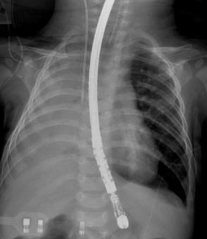
6 month old boy with trisomy 21 weighing 5.6kg presented for VSD repair. Inhalational induction was performed, intravenous access was obtained, and patient was intubated uneventfully with 3.5 cuffed endotracheal tube. Breath sounds were equal bilaterally and adequate tidal volume of 35ml was achieved on the ventilator with the following settings: pressure control ventilation, pressure 15, PEEP 5, rate 25, FiO2 50%. Right radial artery and right internal jugular vein were cannulated under ultrasound guidance. TEE probe was placed. After several minutes, patient’s tidal volumes decreased to 15ml with the initial ventilator settings, and oxygen saturation dropped to the mid 80 percent. Upon auscultation, no breath sounds were heard over the right lung. FiO2 was increased to 100%, patient was manually ventilated, endotracheal tube position was confirmed, and recruitment maneuver was performed. Upon re-ascultation, faint breath sounds were heard over the right lung, but there was no improvement of tidal volume or oxygen saturation. Chest x-ray showed a complete opacification of the right lung with moderate mediastinal shift to the right. At this point, TEE probe was removed leading to improvement in both tidal volumes and oxygen saturation.
There are many causes of intraoperative hypoxemia, and it becomes a challenge to determine the etiology and effectively manage it. Our differential diagnosis included migration of the endotracheal tube, mucus plugging, atelectasis, bronchospasm, pneumothorax, and bronchus compression, which were systemically ruled out. With the chest x-ray showing a complete opacification of the right lung with mediastinal shift to the right, we determined that the hypoxemia was due to the compression of the right main bronchus by the TEE probe. Probe was promptly removed, with improvement of tidal volume and oxygen saturation. Airway obstruction by TEE probe has been reported in the past.1 In a study of TEE examinations involving 1650 children, Stevenson described airway obstruction in 14 patients (1%).2 In another study of TEE examinations involving 200 children with congenital heart disease undergoing cardiac surgery, Sheil described airway obstruction in in 6 patients (3%).3 Airway obstruction by TEE probe is an infrequent complication, especially the degree of obstruction noted in our case with right main bronchus obstruction. TEE is a relatively safe and invaluable diagnostic tool, however complications do occur which may lead to morbidity and mortality if not recognized and treated promptly.
-
 NM-363 Image 1
Top


