
AIR-9
Uncommon association of Tetralogy of Fallot, Absent Pulmonary valve (APV) and Pierre Robin sequence causing severe airway compromise.
Cruz Beltran S, Miller B, Mukkamala S
Emory University, Atlanta, Georgia, United states of america
Tetralogy of Fallot (TOF) with absent pulmonary valve (APV) is an extreme form of tetralogy where pulmonary insufficiency and mild annular stenosis result in massive pulmonary arterial dilatation. Congenital absence of APV is a rare defect and constitutes 3% to 6% of postnatal cases of TOF. Some characteristics include rudimentary cusps of the pulmonary valve, pulmonary regurgitation and a variable degree of dilatation of the main and branch pulmonary arteries.
Here, we present a case of a 12-week-old with Pierre Robin Syndrome, TOF and APV who developed complete effacement of the carina and proximal main bronchi and presented for laryngeal web resection.Â
Clinical Summary
A 12-week-old girl born at 36 weeks GA, with history of Pierre Robin sequence, TOF with APV and right aortic arch was transferred to our institution post birth for airway evaluation due to persistent desaturation. A bedside bronchoscopy demonstrated a laryngeal web with a very small posterior glottic opening, and patient underwent tracheostomy.Â
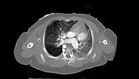
Eight weeks later, the patient presented for laryngeal web resection. At this time, her cardiac condition had progressed to significant enlargement of bilateral pulmonary arteries and mass effect on the carina, which caused near complete effacement of the airway at the carina and proximal right and left main bronchi (Figure).Â
Anesthesia was induced with intravenous fentanyl, rocuronium and sevoflurane via tracheostomy tube. A peripheral IV and arterial line were placed and the patient was maintained on FiO2 0.21%. Patient was placed in suspension laryngoscopy for visualization of the supraglottic structures, which were normal. A grade 3 laryngeal web was identified. The laryngeal web was divided, the upper trachea was normal; however, the mid and distal trachea were severely malacic. The right and left main stem bronchus were malacic as well. She underwent laryngotracheal reconstruction with cartilage grafting. The patient remained stable throughout the surgery. Anesthesia was maintained with sevoflurane and intermittent boluses of short acting opioids. After the tracheal reconstruction, the patient was transferred to the intensive care unit (ICU) for monitoring and mechanical ventilation. Four months later, she remains in the ICU due to chronic respiratory failure and recurrent respiratory infections.Â
Discussion: In this case, we present the rare association of Pierre Robin with TOF, APV, and dilation of the pulmonary arteries with subsequent compression of the tracheobronchial tree. Understanding of the anatomy and patho-physiology is paramount in this case. Induction and maintenance of anesthesia can cause drastic changes in the pulmonary vascular resistance; worsening the obstruction of the lower respiratory tract or triggering serious complications such as a pulmonary hypertensive crisis. Such variations can lead to severe compromise of forward flow and cardiac output.
-
 AIR-9 Image 1
Top


